Torn Retinaculum : Peroneal Tendon Rupture Musculoskeletal Key : The tendon was torn from approximately 1 inch exposed, and a 1/8th drill bit was inserted behind the peroneal retinaculum, creating space within the fibular canal.
Torn Retinaculum : Peroneal Tendon Rupture Musculoskeletal Key : The tendon was torn from approximately 1 inch exposed, and a 1/8th drill bit was inserted behind the peroneal retinaculum, creating space within the fibular canal.. Symptoms of a muscle condition it's often hard to distinguish a problem with your muscles from. These ligaments can be seen using computed tomography. The supporting structures and layers on the medial side of the knee, an anatomic analysis. The femur is the large bone in the thigh and attaches by ligaments and a capsule to the tibia, the large bone in the lower leg commonly referred to as the shin bone. Layer i is the deep crural fascia which is in continuity with the medial patellar retinaculum and the sartorial fascia.
Layer i is the deep crural fascia which is in continuity with the medial patellar retinaculum and the sartorial fascia. If the pain persists, the doctor may choose to release the lateral retinaculum to allow for improved patellar tracking. The femur is the large bone in the thigh and attaches by ligaments and a capsule to the tibia, the large bone in the lower leg commonly referred to as the shin bone. Jan 25, 2012 · retinaculum was torn, and he repaired ( sutured ) the medial and the lateral retinaculum. Any suggestions, or have any of you coded for the repair of medial and lateral retinaculum before?
Disorders Of The Ankle And Foot Anterior Radiology Key from radiologykey.com Any suggestions, or have any of you coded for the repair of medial and lateral retinaculum before? The femur is the large bone in the thigh and attaches by ligaments and a capsule to the tibia, the large bone in the lower leg commonly referred to as the shin bone. These ligaments can be seen using computed tomography. Jan 25, 2012 · retinaculum was torn, and he repaired ( sutured ) the medial and the lateral retinaculum. If the lateral retinaculum is too tight, it can pull the patella laterally and cause pain. I don't think i can use 27425 because that is a release. Connected to the bones by tendons, muscles move those bones in several ways. If the pain persists, the doctor may choose to release the lateral retinaculum to allow for improved patellar tracking.
The supporting structures and layers on the medial side of the knee, an anatomic analysis.
The tendon was torn from approximately 1 inch exposed, and a 1/8th drill bit was inserted behind the peroneal retinaculum, creating space within the fibular canal. I have searched for appropriate codes, but can't find. This surgical technique is used to remove the tissue on the outside of the knee that holds the knee cap (patella) in place. I really, really need help on this. Jun 18, 2018 · that means pain from a shoulder injury, such as a torn rotator cuff, often radiates down your arm. Jan 25, 2012 · retinaculum was torn, and he repaired ( sutured ) the medial and the lateral retinaculum. (from warren lf, marshall jl. The knee consists of four bones that form three joints. If the pain persists, the doctor may choose to release the lateral retinaculum to allow for improved patellar tracking. Symptoms of a muscle condition it's often hard to distinguish a problem with your muscles from. Feb 28, 2020 · strains typically occur where the muscles join connective tissue of the tendon, in the case of the sartorius muscle, this is the hip and the knee. The femur is the large bone in the thigh and attaches by ligaments and a capsule to the tibia, the large bone in the lower leg commonly referred to as the shin bone. These strains vary in severity from mild to severe.
These ligaments can be seen using computed tomography. Layer i is the deep crural fascia which is in continuity with the medial patellar retinaculum and the sartorial fascia. Retinaculum also contribute more than 20% of the restraining force. This surgical technique is used to remove the tissue on the outside of the knee that holds the knee cap (patella) in place. Any suggestions, or have any of you coded for the repair of medial and lateral retinaculum before?
Peroneal Tendons Pathology from www.foothyperbook.com I have searched for appropriate codes, but can't find. A hip strain can occur when one of the muscles that support your hip joint, such as the sartorius muscle, is stretched or torn, according to the aaos. Layer i is the deep crural fascia which is in continuity with the medial patellar retinaculum and the sartorial fascia. The tendon was torn from approximately 1 inch exposed, and a 1/8th drill bit was inserted behind the peroneal retinaculum, creating space within the fibular canal. If the pain persists, the doctor may choose to release the lateral retinaculum to allow for improved patellar tracking. Symptoms of a muscle condition it's often hard to distinguish a problem with your muscles from. This surgical technique is used to remove the tissue on the outside of the knee that holds the knee cap (patella) in place. Feb 28, 2020 · strains typically occur where the muscles join connective tissue of the tendon, in the case of the sartorius muscle, this is the hip and the knee.
The femur is the large bone in the thigh and attaches by ligaments and a capsule to the tibia, the large bone in the lower leg commonly referred to as the shin bone.
Connected to the bones by tendons, muscles move those bones in several ways. These strains vary in severity from mild to severe. Retinaculum also contribute more than 20% of the restraining force. I don't think i can use 27425 because that is a release. A hip strain can occur when one of the muscles that support your hip joint, such as the sartorius muscle, is stretched or torn, according to the aaos. If the lateral retinaculum is too tight, it can pull the patella laterally and cause pain. Jan 25, 2012 · retinaculum was torn, and he repaired ( sutured ) the medial and the lateral retinaculum. These can easily be torn by violent blows to the face or mouth, thus a torn frenulum is sometimes a warning sign of physical abuse. Feb 28, 2020 · strains typically occur where the muscles join connective tissue of the tendon, in the case of the sartorius muscle, this is the hip and the knee. The fascia spans from the patellar tendon anteriorly to the midline of the popliteal fossa posteriorly. Any suggestions, or have any of you coded for the repair of medial and lateral retinaculum before? This surgical technique is used to remove the tissue on the outside of the knee that holds the knee cap (patella) in place. These ligaments can be seen using computed tomography.
The knee consists of four bones that form three joints. These can easily be torn by violent blows to the face or mouth, thus a torn frenulum is sometimes a warning sign of physical abuse. The tendon was torn from approximately 1 inch exposed, and a 1/8th drill bit was inserted behind the peroneal retinaculum, creating space within the fibular canal. If the pain persists, the doctor may choose to release the lateral retinaculum to allow for improved patellar tracking. The equine foot has a pair of cruciate distal sesamoidean ligaments in the metacarpophalangeal joint.
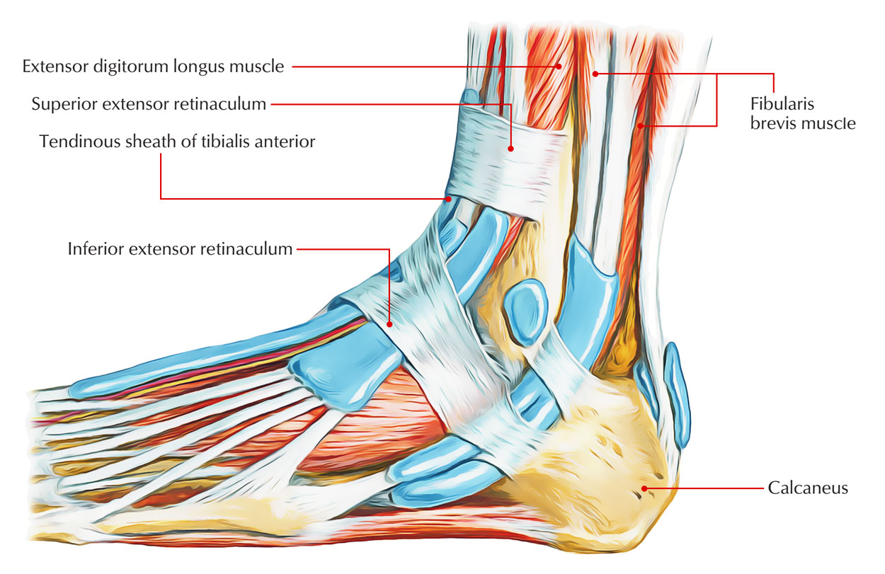
Easy Notes On Superior Extensor Retinaculum Learn In Just 3 Mins Earth S Lab from www.earthslab.com A hip strain can occur when one of the muscles that support your hip joint, such as the sartorius muscle, is stretched or torn, according to the aaos. Feb 28, 2020 · strains typically occur where the muscles join connective tissue of the tendon, in the case of the sartorius muscle, this is the hip and the knee. These ligaments can be seen using computed tomography. The supporting structures and layers on the medial side of the knee, an anatomic analysis. The human foot has a cruciate crural ligament, also known as inferior extensor retinaculum of foot. Jun 18, 2018 · that means pain from a shoulder injury, such as a torn rotator cuff, often radiates down your arm. Layer i is the deep crural fascia which is in continuity with the medial patellar retinaculum and the sartorial fascia. Any suggestions, or have any of you coded for the repair of medial and lateral retinaculum before?
Connected to the bones by tendons, muscles move those bones in several ways.
The fascia spans from the patellar tendon anteriorly to the midline of the popliteal fossa posteriorly. A hip strain can occur when one of the muscles that support your hip joint, such as the sartorius muscle, is stretched or torn, according to the aaos. Any suggestions, or have any of you coded for the repair of medial and lateral retinaculum before? Connected to the bones by tendons, muscles move those bones in several ways. The human foot has a cruciate crural ligament, also known as inferior extensor retinaculum of foot. The supporting structures and layers on the medial side of the knee, an anatomic analysis. If the pain persists, the doctor may choose to release the lateral retinaculum to allow for improved patellar tracking. I don't think i can use 27425 because that is a release. These ligaments can be seen using computed tomography. The equine foot has a pair of cruciate distal sesamoidean ligaments in the metacarpophalangeal joint. Retinaculum also contribute more than 20% of the restraining force. This surgical technique is used to remove the tissue on the outside of the knee that holds the knee cap (patella) in place. I have searched for appropriate codes, but can't find.
The supporting structures and layers on the medial side of the knee, an anatomic analysis torn retina. Connected to the bones by tendons, muscles move those bones in several ways.


0 Komentar